Gold nanoparticle labeling
Detection of viruses is of enormous importance in medical research as well as in clinical diagnosis. Also, detection of virus-like biological particles such as extracellular vesicles (ev) is a topic of intense research and great potential. A great number of detection methods have been devised, ranging from the very quick and simple to the very advanced, but there is still much room for improvement and we have a novel and very interesting method to propose.
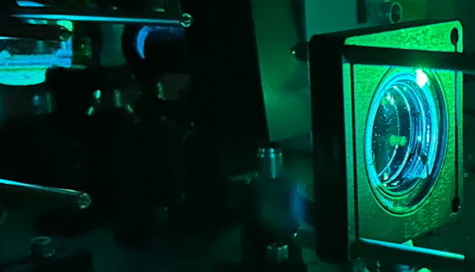
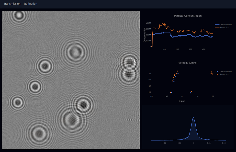
Most methods for virus detection are indirect and involve detecting DNA/RNA or antibodies, or studying the effect of the virus on cells in a cell culture. Molecular-based methods, such as PCR and ELISA and Western blot, can detect residues of DNA/RNA even after an infection has ended, but do not accurately measure the concentration of intact virus particles in a sample. Electron microscopes can count virus particles, but require expensive equipment and extensive training to use. Fluorescent dyes combined with analytical techniques like NTA, fluorescence microscopy and flow cytometry can also be used to detect viruses and ev, but many fluorescent dyes rapidly bleach under laser illumination, making analysis more difficult. In complex biological samples such as blood serum which contains much protein and particles, fluorescent dyes can cause too much background signal by attaching to all the other material in addition to the targeted virus or ev. Our innovative and patent-pending method to address these shortcomings is to label viruses with nanoparticles of gold rather than fluorescent dyes, and to detect them selectively with our holographic nanoparticle tracking method. The concept is based on the fact that holographic nanoparticle tracking is very good at differentiating between strongly light absorbing particles such as gold nanoparticles and weakly light absorbing particles such as biological nanoparticles.
The advantages are the following:
- Selectively detecting only virus particles with gold particles attached, whereas individual virus and gold can be kept below detection limit and not disturb the measurement. This makes use of that holographic particle tracking is insensitive to particles below detection limit, which blend into the background.
- Particles above detection limit, but without gold nanoparticles attached, are readily differentiated from those with gold attached.
- Comparably few gold nanoparticles need to be attached to enable detection, whereas with most analytical methods many fluorescent molecules would be needed. This can both enhance detection probability and enable detection of binding sites of which there are only few per virus/ev particle.
- Combined with size measurement, intact virus/ev particles can be differentiated from virus/ev debris.
- Gold nanoparticle labeling enables detection of smaller virus/ev particles than can be detected without labeling.
- Gold nanoparticles are not bleached by intense laser illumination.
The concept was first demonstrated by Olsén, E, et al 2023, and further research is ongoing. We are interested in collaborations to develop and evaluate this method further. Please contact us if interested!