Extracellular Vesicles

See more, clearly.

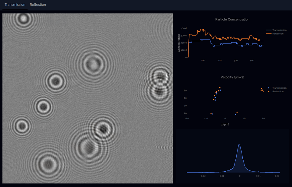
DAISY analysis is perfectly suited to rapid and convenient characterisation of extracellular vesicle samples. Alongside accurate size and concentration quantification, DAISY reveals refractive index and geometry details of your particles. This allows you to discern extracellular vesicles from potential contaminants, such as lipoproteins and protein aggregates, with ease.  All of this information can be gathered irrespective of the liquid in which your sample is contained. 

What are extracellular vesicles and exosomes?

Extracellular vesicles are nano-sized vesicles secreted into the extracellular environment by a host of cell types. Exosomes represent a particular class of small (50-150 nm) extracellular vesicles produced within the endosomal system and released via organelle fusion with the plasma membrane. As well as exosomes, extracellular vesicles can include microvesicles, derived from budding of the plasma membrane, and the largest extracellular vesicle class, apoptotic bodies, arising from cells undergoing programmed cell death.  

Biological function

Extracellular vesicles have been shown to contain a wide range of potentially bio-active molecules such as DNA, RNA, proteins, and metabolites. As a result of their cargoes and their extracellular nature, the idea of extracellular vesicles functioning in inter-cellular communication has growing support. Extracellular vesicle-mediated communication has been implicated in a wide range of physiological processes and disease associated pathophysiological states. Some key areas of particularly active research are:

  • Cancer research
  • Cardiovascular disease
  • Immunology
  • Stem cell paracrine function

Health-tech and biotech applications

The discovery that extracellular vesicles contain biomolecules, effectively protecting them from degradation, has spurred huge and growing interest in their potential applications. Broadly, the interest in extracellular vesicles falls into three areas, diagnostics, extracellular vesicle-based treatments, and harnessing extracellular vesicles as therapeutic delivery vectors. 

 Extracellular vesicles have several key advantages when it comes to diagnostic applications. They are enriched in bodily fluids such as urine compatible with minimally invasive liquid biopsy approaches. Through encapsulating and protecting bio-molecules, extracellular vesicles contain a detailed picture of cellular status of their parent cell. In the case of RNA and DNA, sequencing approaches can reveal specific diagnostic markers for disease such as the presence cancer mutations. Additionally, their membrane-bound nature makes extracellular vesicles relatively easy to enrich for example via centrifugation or filtration. Demonstrating the promise of extracellular vesicle-based diagnostics, tests are already in clinical use and showing real benefits over existing tests in the detection of prostate cancer. Further work in identifying extracellular vesicle associated biomarkers is underway and developing at a rapid pace.  

Beyond diagnostics, the application of extracellular vesicles as direct therapeutic agents has some support. In particular, mesenchymal stem cell derived extracellular vesicles have been posited to be the driving force behind the beneficial effects seen with stem cell therapies. 

 Another key application of extracellular vesicles is in the delivery of therapeutic molecules. This approach relies on the biocompatibility, cargo-protective effects, and potential for cell type specific targeting that are inherent in extracellular vesicles. There is also a growing interest in engineered “hybrid EV” systems incorporating the cellular delivery capabilities of engineered lipid nanoparticles and liposomes and the targeting capacity of viral envelope proteins. 

 Although a relatively young field, it seems clear that extracellular vesicle research has huge potential.

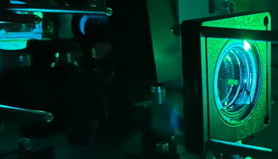
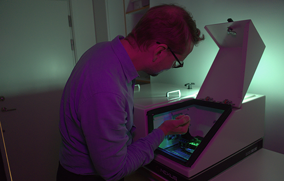
 Characterisation of extracellular vesicles by DAISY NTA

One key problem in working with extracellular vesicles is that their small size makes in depth characterisation and comparison between samples difficult. This invisible sample heterogeneity can lead to apparent variability in effects, which could likely be explained with a better understanding of the precise composition of the extracellular vesicle samples. It is in providing this in-depth characterisation that DAISY analysis excels. Standard approaches to characterisation such as nanoparticle tracking analysis (NTA), dynamic light scattering (DLS), and electron microscopy (EM) are each able to provide estimates of particle size, alongside some other sample characteristic such as concentration (NTA), polydispersity index (DLS, EM), particle geometry (EM). DAISY analysis is unique in that it can give accurate particle size, concentration, geometry, and refractive index, all in a single analysis. This multi-dimensional information can be used to identify contaminants, aggregates, and deviations from reference samples on a sample by sample basis with ease.

What is DAISY analysis?

The DAISY instrument captures video recordings of particles flowing through a microfludic channel. The video recordings are subsequently analyzed to extract information about the particles, both from their Brownian movement and from their optical properties. ?

The instrument illuminates the sample from two opposite directions, and the light from these two directions is recorded in two different video recordings. The advantage of the dual-angle illumination is both to enhance the particle size range, and to extract more optical information from the particles.

What can DAISY tell me about my sample?

  • Concentration
  • Size distribution (from 50 to 1000 nm)
  • Refractive index
  • Geometry         

Key advantages of DAISY analysis

  • Label-free
  • Ability to distinguish EV from other contaminating particles, such as lipoproteins and protein aggregates
  • Fast and accurate (from 20 seconds per sample of data acquisition)
  • Independent of sample media composition (optical sizing, viscosity)
  • Low sample requirements (from 10 µl per sample)

Features & benefits

  • Hydrodynamic diameter
  • Optical diameter
  • Refractive index
  • Morphology/shape
  • Particle density
  • Concentration
  • Size range 50nm – 5 µm
  • Single-use microfludic chips
How it is used

How it is used

Analysis of particles in liquids is of great importance in many fields, ranging from life science to water analysis.

Read more

How it works

When highly ordered laser light passes through the sample, it collects detailed information which is extracted digitally using advanced image processing and machine learning.

Read more

About us

Holtra was founded in 2020, based on cutting-edge research at Chalmers and Gothenburg Universities, and is currently developing its first analytical instrument.

Read more